ID22 Bio Nano Crystallography (BioNX)
1. Introduction
The Bio Nanocrystallography (BioNX) beamline is the first designated MX beamline for the Korea-4GSR during its initial phase. High coherence and small beam divergence with high photon flux are advantages of the 4th generation synchrotron, which enable the design of nanocrystallography beamline. The in-vacuum undulator20 (IVU20) serves as the light source, and the beam is tunable within the energy range of 8-25 keV. A horizontal Double Crystal Monochromator (hDCM) is chosen to achieve the required beam stabilization. The targeted beam size at the sample position ranges from 1x1 to 13x13 μm2. Maximum focusing of the beam will be achieved using a Vertical Focusing Mirror (VFM) and a Horizontal Focusing Mirror (HFM). Compound refractive lenses (CRLs) will be used to adjust the beam size as needed to accomplish the fast beam expansion. At the end station, the MD3 diffractometer and EIGER2 XE 16M detector, along with a high-capacity sample changer, will be installed. This beamline is designed to determine the crystal structure of proteins ranging from nano to micro-sized crystals. The experiments to be implemented on this beamline include serial synchrotron crystallography, in-situ crystallography, automated high-throughput screening, and X-ray fluorescence (XRF). These techniques will enable comprehensive structural and functional analysis of biological macromolecules, facilitate efficient drug discovery, and provide detailed elemental composition data. Additionally, a high-throughput fragment screening facility will be established for drug discovery experiments.
2. Scientific objective
2.1 Serial synchrotron crystallography
The primary technique of the BioNX beamline is serial synchrotron crystallography (SSX). The demand for analyzing the crystal structures of challenging proteins has recently increased. Determining crystal structures using nano to micro-sized protein crystals obtained from limited protein quantities has become an essential technique in the field of structural biology. This approach is particularly valuable for studying proteins that are difficult to crystallize in larger sizes, thus enabling the structural elucidation of complex and scarce proteins. To achieve this, serial crystallography techniques have been developed to reduce radiation damage with high photon flux and small beam size of the developed synchrotron source. To meet this demand, BioNX beamline will be designed to utilize a 1-micron beam and a photon flux exceeding 10^14 photons per second. In addition, not only an MD3 diffractometer with a sphere of confusion value below 200 nm for precise sample alignment but also an EIGER2 XE 16M detector, capable of capturing images at up to 560 Hz in full frame mode will be established. This combination will deliver a high-performance SSX system, enhancing the efficiency and accuracy of the data collection. The sample delivery system will primarily provide fixed-target, with plans to implement the liquid jet method.
2.2 In-situ crystallography
In-situ crystallography is a technique used to study the structural properties of crystals directly in their native environment, without the need for crystal extraction or transfer. This method minimizes potential damage and artifacts that can arise during handling and exposure to non-native conditions. By maintaining the crystals in their natural state, in-situ crystallography provides more accurate and reliable data on the molecular structure and dynamics of the sample. This approach is particularly useful for studying fragile and difficult-to-crystallize proteins and other macromolecules. The state-of-the-art BioNX beamline, combined with the small-wedge synchrotron crystallography (SWSX) technique and advanced computing power, will provide user-friendly access to in-situ crystallography experiments.
2.3 Automated high-throughput screening
Automated high-throughput screening using MX beamlines at synchrotrons has significantly accelerated the identification of potential drug candidates by rapidly analyzing numerous protein-ligand complexes. This is achieved by automating sample handling and data collection, which facilitates the efficient screening of large compound libraries. The BioNX beamline will support remote sample delivery and experimentation, enabling the screening of over 600 samples per day. Additionally, a dedicated high-throughput fragment screening laboratory will be established, allowing users to conduct experiments directly at the accelerator facility. This approach not only enhances the speed and precision of drug discovery but also provides detailed insights into the binding interactions and mechanisms of action of potential therapeutic agents, thereby improving the overall drug development process.
3. Beamline Requirements for the Insertion Device
The BioNX beamline employs the in-vacuum undulator 20 (IVU20), which delivers a highly coherent flux and a low heat load of 12 kW (Table 1). However, it has a feature of a spectral discontinuity between 6.8 keV and 8.08 keV, thereby limiting the detection of nickel and cobalt during XRF experiments (Figure 1). The BioNX beamline will cover an energy range of 8-25 keV with primary operations planned at energy levels of 12 keV and 20 keV.
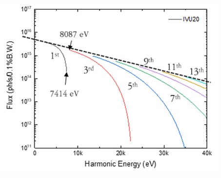
Figure 1. Predicted photon flux of the IVU 20
Table 1. Source Parameter for BioNX beamline
Undu lator |
Period [mm] |
Length [m] |
Location |
Max Power |
IVU20 |
20 |
3.0 |
Center of Straight section |
1.15 kW |
Maximum power refers to the maximum beam power passing through a mask with an aperture of 2 x 2 mm² @ 23 m using the IVU20.
4. Beamline Requirements for Front End
The front end of the BioNX beamline will employ the latest High Heat-Load Front End, capable of handling up to 12 kW of power designed to accommodate the in-vacuum undulator 20 (IVU20) beam. This front-end system will transmit the central cone beam with a divergence of 2.4 sigma (23 μrad). Further details are provided in the ‘Front End’ section of this report.
5. Beamline Layouts
The layout of the BioNX beamline consists of a front-end, an optical hutch, a long spool, and an experimental hutch (Figure 1). Two separate enclosed hutches, the optical hutch and the experimental hutch, will be operated and located at sector ID22. The distance from the undulator to the sample is 70 meters (Figure 2). Table 1 presents all major beamline components and their respective locations along the beamline.
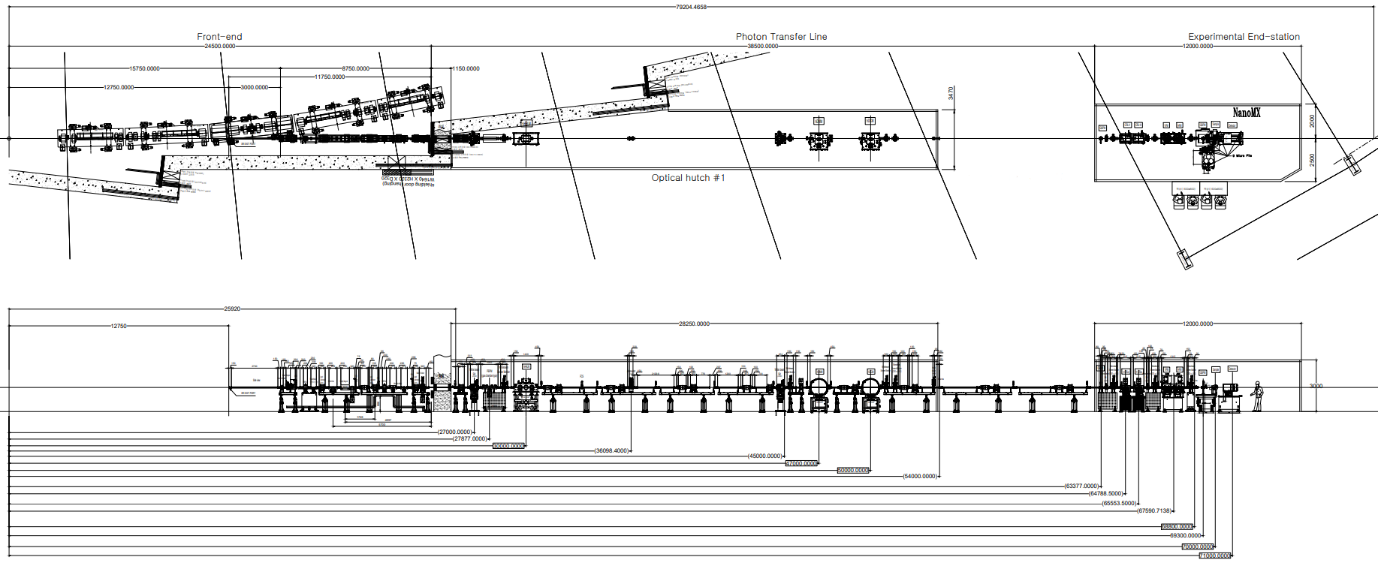
Figure 2. Beamline layout of BioNX
5.1 Beamline Component Table
Table 2. Component list of BioNX beamline
Hutch distance (m) |
Distance (m) |
Component |
Specification |
Description |
|---|---|---|---|---|
Optical Hutch 26 – 52 m |
27 |
White beam slit |
4-way slit, UHV |
White beam conditioning |
30 |
High heat-load mirror |
Flat mirror 2-stripe coating (Pt, Rh) |
Reduce heat load |
|
36 |
Attenuator |
CVD Diamond |
Reduce heat load |
|
45 |
White beam slit |
4-way slit, UHV |
Beam conditioning |
|
46 |
White beam screen monitor |
Retractable; CCTV Diamond BPM |
White beam monitor |
|
47 |
horizontal DMM (1) |
Ru/B4C |
M onochromator Pink beam |
|
50 |
horizontal DCM |
Si(111) crystal |
M onochromator Monobeam |
|
51 |
White beam screen monitor |
Diamond BPM |
White beam monitor |
|
52 |
Monobeam screen monitor |
CCTV; cooled |
Monobeam profiling |
|
End-station 65-75m |
65 |
QBPM |
Ultra High Vacuum QBPM (UHV QBPM) Feedback |
Beam position & profile defining |
66 |
Monobeam Slits |
4-way slit, UHV |
Beam conditioning |
|
66.2 |
Monobeam screen monitor |
CCTV; cooled |
Monobeam profiling |
|
66.5 |
CRL-vertical |
1D, Be coating |
Beam size expansion |
|
67 |
CR L-horizontal |
1D, Be coating |
Beam size expansion |
|
67.2 |
Monobeam Slits |
4-way slit, UHV |
Beam conditioning |
|
67.5 |
Monobeam Sceen monitor |
CCTV; cooled |
Monobeam profiling |
|
67.7 |
VFM |
Si/ Rh (50) /Pt (50) |
Focusing vertical beam |
|
68.4 |
HFM |
Si /Rh (50) /Pt (50) |
Focusing horizontal beam |
|
68.6 |
Monobeam screen monitor |
CCTV; cooled |
Monobeam profiling |
|
69.3 |
QBPM |
High Vacuum QBPM (HV QBPM) |
Beam position & profile defining |
|
70 |
Sample stage |
MD3-UP SOC: <200 nm |
Beam di ffractometer |
|
~71 |
Detector |
EIGER 2 XE 16M |
Detection of diffraction pattern |
Under consideration